


Introduction
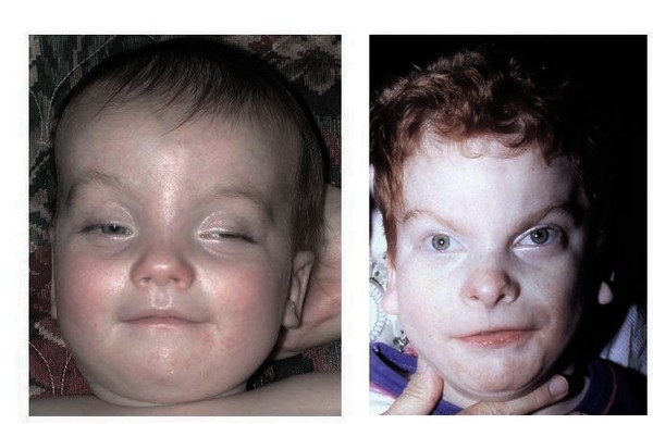
The Miller-Dieker Syndrome (MDS) is a rare genetic brain disorder attributable to chromosomal defect, which results in malformation of crucial parts of the brain. In patients with MDS, the exterior of the brain is characterized by microcephaly and lissencephaly, meaning that the brain is smaller and the neocortex is abnormally smooth with fewer grooves and folds, respectively, compared to a normal brain. MDS is caused by a deletion at a region of chromosome 17 responsible for neural development. In addition, patients with MDS present with severe intellectual disabilities and seizures, and most patients do not survive beyond infancy.
Impact of MDS on family
In some cases, people with MDS inherit the abnormal chromosome from a parent who is unaffected. However, in other cases, the deletion of the genes attributed to MDS happens randomly during early fetal development or in reproductive cells. Typically, individuals affected do not have a history of MDS in their families. The disorder is characterized by numerous health problems including severe seizures, and their quality of life is also severely affected. People with MDS require extensive supportive care as a result of poor muscle coordination, lack of tendon reflexes, muscle weakness, and respiratory problems. In this case, MDS might put a strain on families owing to the need for consistent care and support.
Aim, design and findings of the study
The Human Brain “organoids” Offer New Insight into Rare Developmental Disease (2017) study aimed to investigate the development of MDS using brain organoids of the developing human brain. To accomplish this, the scientists grew 3D organoids in culture dish, creating a form of “mini brains”. These models offer valuable insight into the processes of cell differentiation into complex tissues. The use of mouse models to study Miller-Dieker is limited owing to the natural smoothness of the mouse brain. As patients with MDS present with a smooth outer layer of the brain, a mouse model to establish the genetic process behind the smoothness would be flawed. The mouse brain also lacks outer radial glia, which facilitate the expansion of the brain in both complexity and size in primates. Therefore, the are significant differences in growth and development between the human and mouse brain. Furthermore, there are numerous neural stem cells in humans that are absent in mouse.
The Extract in the Context of Miller-Dieker syndrome.
The extract on MDS provides important information on breakthroughs in the study of rare genetic disorders such as MDS. Furthermore, scientist would be better placed to identify the specific genes deleted during brain development and the exact neural stem cells that are altered in MDS, providing further insights into early intervention alternatives for infants with MDS. In addition, the connection in the 3D tissue structure cannot be identified in 2D cell culture models or in animal models. The research team presented novel findings regarding neuroepithelial cells in MDS; the cells die at high rates, and those surviving divide abnormally. Such insight into how the early stages of brain development are altered in people with MDS is crucial to the understanding of lissencephaly of MDS brains.