Diagnosis and determination of Ebola virus titre that was gotten from a healthcare worker deployed in the selected West African country; Zaire
Introduction
Ebola virus has led to high mortality rate and dire human race together with disruption of the economy within West Africa. The disease has continuously shown an ominous threat within the region and other parts of the world. It has put at risk the provision of healthcare together with other critical services within these regions. The rate at which the disease has affected the health system has resulted in increased scientific studies on the diagnostic, treatment, and prevention of the disease via upsurge available aid. Currently, the advanced knowledge of the virus pathogenesis, risk-factor, transmission mode, control, and sociocultural factor has dramatically increased within our environment (Ansari, 2014).
In this study, a female healthcare worker within the selected West African countries such as DRC of age forty-five was discovered to have haemorrhagic fever-like symptoms in August 2020. These symptoms exhibited were fever, malaise, and continuous vomiting for twelve days after she came back from the United Kingdom. After the exhibition of these symptoms from the health officer, this patient was admitted after being isolated from her area of duty in London. (Cooper et al., 2018).
Diverse samples from serum, saliva as well as CSF were taken and taken for culture in the Biosafety level four located in the Porton Down to examine the virus employing PCR thoroughly, and the virus was cultured in a baby hamster kidney cells to comprehensively determine the hemorrhagic fever diseases such as the Zaire Ebola viruses as well as estimation of the viral tire in every sample (Cooper et al., 2018).
During the laboratory activities’ initial session, viral DNA was extracted from the clinical sample before the actual polymerase chain reaction process.
Additionally, the clinical specimen obtained from the saliva, serum, and CSF from the patient was then infected to the baby hamster kidney cells and seeded in ninety-six well plate to propagate the virus from the sample effectively. In the second session, the virus titer was then determined in every piece collected. Full agarose gel electrophoresis was conducted to visualize the precise virus PCR-amplicon within the clinical model and comprehensively analyze the sequence information obtained PCR-amplicon and then fully sequenced (Warfield et al., 2003). Visualization of this virus can be done through an electron microscope; it is observed in the studding virion region as a filamentous viral particle with protein molecules and its cell surface structure. In this region, the virus undergoes development, referred to as the budding region, seen on the microscope. To amplify the cDNA amplicons of this virus, we use the RT-PCR technique (Li et al., 2015). In this process, the biochemical test carried out on the patient investigates the serum samples and tries to determine if the virus has reached the acute salivary stage on the named patient. This virus can also be explored through the use of the immunohistochemical staining technique. This technique takes a small sample of tissue, and it is then visualized under a microscope. If the example is positive for Ebola, there will be brown staining (Cooper et al., 2018).
The Ebola virus genus is non-segmented, negative-sense, and have a single-stranded RNA. The virus belongs to the Filoviridae family, a word generated from a Latin name known as filum to mean a threadlike structure. This virus has varying molecular shapes for its specific elements of their RNA structural molecules; this allows the lipid bilayer to form new systems in the surface of the cells found in the host tissues, other viruses in this family include the Marburg virus was discovered in Germany. These viruses have similar genomic sequences, same replication methodology; they also belong to the same family of rhabdoviruses and paramyxoviruses (Onyango, 2007).
Ebola virus genome composed of single-stranded RNA estimated to have nineteen thousand nucleotides long. Often, the virus genome consists of seven well-known nucleotide sequences, which code for the structural and non-structural proteins called viral proteins, i.e., VP. Ebola virus core structure is composed of RNA genomic molecules consisting of nucleoprotein (Ansari, 2014). Typically, viral proteins consist of diverse types of viral proteins, with each having various functions. Viral proteins such as VP 30 often assist in activating the RNA transcription process of the virus that highly rely on the concentration of the VP30. VP24 is often known as the core matrix protein and also the most abundant virion element of the virus (DeMers, 2020).
Viral protein 35 assists in synthesizing RNA of the virus and thus acts as the interferon antagonist. Naturally, there is a likelihood that the potency level of VP35 can effectively account for the diverge degree of virulence amid the diverse strain of the Ebola virus. Viral protein 40 serves as the matrix protein from the negative strand of the virus RNA. VP40 assists in the assemblage of lipid enveloped virus by providing linkage amid the surrounding membrane and the nucleotide structure (Liu, et al., 2015). Additionally, the protein can also be termed as single surface transmembrane glycoprotein leading to the formation of spikes on virion and the achievement of viral entry into the body cells through mediation receptor binding, fusion as well as precise target body cell entry (Liu, et al., 2015).
Methodology
Section 1
In this section, the following practices were carried: Titration of the virus was gotten from hospital samples that were collected from one of the health officers in a BHK cell line; secondly, the RNA extracted from the virus were isolated, thereafter, they were amplified using RT-PCR technique. This was done to reserve the protein molecules of the virus.
Dilution was done for the clinical samples that were obtained from the patient, these samples included the blood, mouth wash and body fluids. RNA was extracted from these samples. The BHK cell lines were infected also. Lastly, the determination of virus sensitivity of two strains of Influenza H1N1 to an antiviral compound using the plaque reduction assay.
Section 2
This was the second part of the practical where X-gal was used to stain the cell lines in the clinical specimens. This was done to the viral titre as well. The results of the RT-PCR samples were eventually analysed and a study of the sequences done.
Results
From the outcome below ß galactosidase gene is used as a reporter gene and X-Gal- generates indigo-blue color in cells which express ß galactosidase
Figure 2
50ul 25ul 12. 5ul 6.25ul 3.13ul
Figure 3
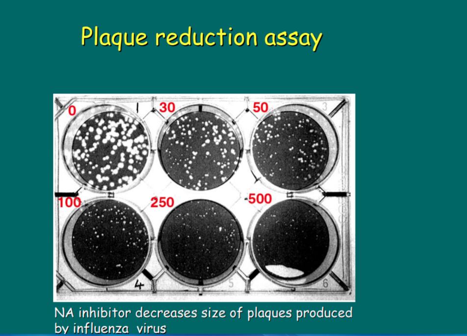
The figure below shows the infections of the individual particles of the virus. This data was generated from specific set up protocols that were used during the practical. The plaques in the assay were identified either as areas of dead/destroyed cells detected by general cellular stains or as areas of infected cells detected by immuno-staining
The sequence of the Ebola virus obtained from the CFS result are stated below;
>TTGAAATTTATATCGGAATTTAAATTGAAATTGTTACTGTAATCACACCTGTTTTGTTTCAGAGCCATGT
CACAAAGATAGAGAACAACCTAGGTCTCCGAAGGGAGCAAGGGCATCAGTGTGCTCAGTTGAAAATCCCT
TGTCAACATCTAGGTCTTATCACATCACAGGTTCCACCTCAGGTTCTGCAGGGTGATCCAACAACCTTAA
TAGAAACATTATTGTTAAAGGACAACATTAGGTCACAGTCAAACAAGCAAGATTGAGAATTAACCTTGAT
TTTGAACTTGAACACCTAGAGGATTGGAGATTCAACAACCCTAAAGCTTGGGGTAAAACATTGGAAATAG
TTAAAAGACAAATTGCTCGGAATCACAAAATTCCGAGTATGGATTCTCGTCCTCAGAAAGTCTGGATGAC
GCCGAGTCTCACTGAATCTGACATGGATTACCACAAGATCTTAACAGCAGGTCTGTCCGTTCAACAGGGG
ATTGTTCGGCAAAGAGTCATCCAAGTGTATCAAGTAAACAATCTTGAGGAAATTTGCCAACTTATCATAC
AGGCCTTTGAAGCAGGTGTTGATTTTCAAGAGAGTGCGGACAGTTTCCTTCTCATGCTTTGTCTTCATCA
TGCGTACCAAGGAGATTACAAACTTTTCTTGGAAAGTGGCGCAGTCAAGTATTTGGAAGGGCACGGGTTC
CGTTTTGAAGTCAAGAAGCGTGATGGAGTGAAGCGCCTTGAGGAATTGCTGCCAGCAGTATCTAGTGGAA
AAAACATTAAGAGAACACTTGCTGCCATGCCGGAAGAGGAGACGACTGAAGCTAATGCCGGCCAGTTTCT
CTCCTTTGCAAGTCTATTCCTTCCGAAATTGGTAGTAGGAGAAAAGGCTTGCCTTGAGAAGGTTCAAAGG
CAAATTCAAGTACATGCAGAGCAAGGACTGATACAATATCCAACAGCTTGGCAATCAGTAGGACACATGA
TGGTGATTTTCCGTTTGATGCGAACAAATTTCTTGATCAAATTTCTCCTAATACACCAAGGGATGCACAT
GGTTGCCGGGCATGATGCCAACGATGCTGTGATTTCAAATTCAGTGGCTCAAGCTCGTTTTTCAGGTTTA
TTGATTGTCAAAACAGTACTTGATCATATCCTACAAAAGACAGAACGAGGAGTTCGTCTCCATCCTCTTG
The sequence obtained from serum is;
>TTGAAATTTATATCGGAATTTAAATTGAAATTGTTACTGTAATCACACCTGTTTTGTTTCAGAGCCATGT
CACAAAGATAGAGAACAACCTAGGTCTCCGAAGGGAGCAAGGGCATCAGTGTGCTCAGTTGAAAATCCCT
TGTCAACATCTAGGTCTTATCACATCACAGGTTCCACCTCAGGTTCTGCAGGGTGATCCAACAACCTTAA
TAGAAACATTATTGTTAAAGGACAACATTAGGTCACAGTCAAACAAGCAAGATTGAGAATTAACCTTGAT
TTTGAACTTGAACACCTAGAGGATTGGAGATTCAACAACCCTAAAGCTTGGGGTAAAACATTGGAAATAG
TTAAAAGACAAATTGCTCGGAATCACAAAATTCCGAGTATGGATTCTCGTCCTCAGAAAGTCTGGATGAC
GCCGAGTCTCACTGAATCTGACATGGATTACCACAAGATCTTAACAGCAGGTCTGTCCGTTCAACAGGGG
ATTGTTCGGCAAAGAGTCATCCAAGTGTATCAAGTAAACAATCTTGAGGAAATTTGCCAACTTATCATAC
AGGCCTTTGAAGCAGGTGTTGATTTTCAAGAGAGTGCGGACAGTTTCCTTCTCATGCTTTGTCTTCATCA
TGCGTACCAAGGAGATTACAAACTTTTCTTGGAAAGTGGCGCAGTCAAGTATTTGGAAGGGCACGGGTTC
CGTTTTGAAGTCAAGAAGCGTGATGGAGTGAAGCGCCTTGAGGAATTGCTGCCAGCAGTATCTAGTGGAA
AAAACATTAAGAGAACACTTGCTGCCATGCCGGAAGAGGAGACGACTGAAGCTAATGCCGGCCAGTTTCT
CTCCTTTGCAAGTCTATTCCTTCCGAAATTGGTAGTAGGAGAAAAGGCTTGCCTTGAGAAGGTTCAAAGG
CAAATTCAAGTACATGCAGAGCAAGGACTGATACAATATCCAACAGCTTGGCAATCAGTAGGACACATGA
TGGTGATTTTCCGTTTGATGCGAACAAATTTCTTGATCAAATTTCTCCTAATACACCAAGGGATGCACAT
Discussion
In section 1, serial dilution was done to reduce the number of viral cells. This eases the identification of the cell lines under the microscope. During the determination of the virus concentration, a transmission electron microscope is used to determine the viral particle concentration in each sample obtained from the patient (Kreuels, 2014). The number of the particles is therefore calculated in mL following the number of PFU.
In this practical, galactosidase gene used as a reporter gene, and X-gal generates indigo-blue color in cells that express galactosidase. This assay was used to determine the titer of Ebola dose titrations that were done. A control experiment can be set using animal samples; the day of the animal exposure to the virus must be determined to achieve accuracy in the experimental setup. Eventually, deep sequencing of the extracted RNA is done in addition to the sample separation. To increase the depth of the material’s sequence, the mRNA, rRNA, and DNA should be removed from the sample. This process is achieved through cleaning of the pieces before taking them for sequencing. QIAprep mini rep (QIAGEN) can be used to clean these samples before sequencing.
Effective detection as well as confirmation of the viral hemorrhagic-fever outbreak in most of the regions across the globe, mainly West Africa has made it hard for most of the health workers to obtain suitable clinical specimen from the target audience. Precisely, most of the needed requirement for effective collection of samples such as blood or serum has led to vital delay of the notification as well as the whole process of diagnosis of the filovirus virus cases within most regions. Due to absence of available sampling tools, and weak infrastructure within both communication together with transportation and lack of proper trained experts, all these factors has led to delay and inappropriate results. Moreover, other factors such as cultural objection to the collection of blood sample and postmortem invasive sample collection are also attributed and seen as critical factors hindering the entire process. Majority of these factors are experienced within the prevalent regions such as Africa.
The aspect of reluctance within the entire process of permitting invasive techniques used to obtain the clinical specimen often resulted in the querying of whether the non-invasive techniques used to obtain the sample such as oral fluids will be suitable. Samples such as oral fluids like saliva often provide numerous merits over serum method collections. Typically, this is often linked to the reasons such as these techniques are non-invasive, safe and absence of cultural barriers
Ebola attacks the blood vessels; it has a higher concentration in the blood; this is why several dilutions are needed to detect the blood serum. This explains why it was present at a lower serial dilution value as compared to the CSF samples.
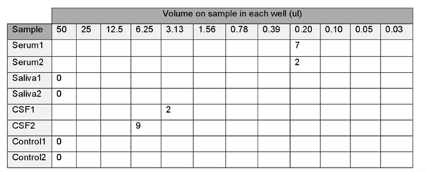
Table showing the number of cells seen under the microscope in different volumes of titer
From these results, in serum one at 0.20µl, seven viral cells were observed. Serum 2 indicated two viral cells at 0.20µl well. CSF 1 shown positive results at 3.13µl well, with two viral cells being observed. CSF 2 shown nine viral cells at a 6.25µl well plate. Finally, controls 1 and 2 did not show any positive results for viral cells in any of the wells.
CSF samples showed positive results at a higher titer value as compared to the serum samples.
In the plaque assay practical, every infectious virus was observed to be multiplying under specialized conditions that result in a localized area of the infected cells or plaque. The plaques assays were honored to be dead while some had destroyed cells identified by general cellular strains or regions of the infected cells detected by immune-staining.
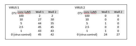
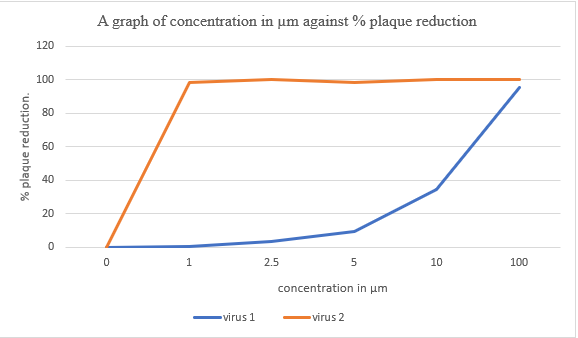
The following sensitivity of the two strains of influenza H1N1 to an antiviral compound was obtained from the results obtained. The averages of the viral results were calculated for individual virus in their respective concentration, and then the % reduction was calculated using the following formula:
100 – (mean of test wells ÷mean of control wells x 100)
For virus 1;
@100µm = 100 – (2/ 43.5 x 100) = 95.40%
@10µm = 100 – (28.5/ 43.5 x 100) = 34.48%
@5 µm = 100 – (39.5/43.5 x 100) = 9.20%
@2.5 µm = 100- (45/43.5 x100) = 3.45%
@1 µm = 100 – (43/43.5 x 100) = 0.46%
For virus 2;
@ 100 µm = 100 – (0/25.5 x 100) = 100%
@ 10 µm = 100 – (0/25.5 x 100) = 100%
@ 5 µm = 100 – (0.5/25.5 x 100) = 98.04%
@ 2.5 µm = 100- (0/25.5 x 100) = 100%
@1 µm = 100 – (0.5/ 25.5 x 100) = 98.04%
Table showing concentration of the viral cells for viruses 1 and 2 in µm and % plaque reduction.
| Virus 1 | Virus 2 | |||
| Mean plaques | % plaque reduction | Mean plaques | % plaque reduction | |
| 100
|
2 | 95.40 | 0 | 100 |
| 10 | 28.5 | 34.48 | 0 | 100 |
| 5 | 39.5 | 9.20 | 0.5 | 98.04 |
| 2.5 | 45 | 3.45 | 0 | 100 |
| 1 | 43 | 0.46 | 0.5 | 98.04 |
| 0 |
During the identification of virus cultures in the lab, we recommend that we use PCR because this procedure is specific and sensitive for non-culturable viruses; it requires less time to detect the pathogen. Viral cultures are used. The viral culture gives a director measure of infectivity, large sample volume (100µm). However, these methods pose the following challenges during the study: they require a more extended period for detection (different viruses have different time variations for detection). The cell culture does not detect the slow-growing and non-culturable viruses. PCR does not determine the infectivity of the viruses being investigated.
During practicals, a lot of care must be taken to avoid contamination of the virus, and Ebola is a viral hemorrhagic fever of humans. Therefore, the extraction must be undertaken under BSC 4; Qiagen sample and assay technology kit is preferred for extraction of this virus as it gives pure DNA, the thermocycler machine should be cleaned and disinfected using 15% ethanol to remove impurities, PCR conditions should be appropriately set as the virus-specific PCR conditions of RT temperature of 50oC for 30m minutes, initial melt 0f 950C for 15 minutes, denaturation temperature of 940C for 1 minute, the annealing temperature of about 500C to 680C for one minute, there should be extension temperature of 720C and the final extension temperature of 720C should run for ten minutes maximum, 40 cycles are run for this set up. DNA ladder is used when running the agarose gel to assist in reading the samples’ base pairs; it should be able to disintegrate appropriately. Correct voltage should be used to run the gel. As seen in this setup, the introduction of viral gene therapies helps us evaluate various body infections. This strategy assists scientists in targeting some abnormalities in the body. These create a new design in therapeutic interventions. Ebola virus, as a result, is always used to estimate viral tire in human samples in hospitals and several research institutions all over the world.
The following techniques can be used in viral detection in the cultivation of viruses: immunoblotting, hemagglutination assay, immunoprecipitation, viral flow cytometry, and, lastly, immunofluorescence assay. To treat Ebola, the appropriate medication should be offered to support blood pressure, vomiting, diarrhea, fever, and pain; this can be achieved by treating other infections in the body. Intravenous injection of body salts is conducted to boost body fluids Warfield et al., 2003.
Lastly, oxygen therapy is conducted on the patients; this boosts and maintains their oxygen level. In small animals, BCX4430 has been proven to be useful for treating small livestock animals (Kreuels, 2014). To control Ebola, contact with the primates such as chimpanzees and monkeys should be minimized, hunters and gatherers should be discouraged from taking bushmeat. Funerals that involve Ebola suspects should be avoided. World Health Organisation recommends home-based treatment to prevent the disease’s spread to the health care workers and the public (Warfield, et al., 2003).
Typically, the RT polymerase chain reaction can be termed as the best technique to detect the Ebola virus as indicated from the result, due to the consistent outcome with all the oral body fluids samples. The reverse polymerase chain reaction method uses diverse oral fluids specimen. Therefore, from the result one can effectively deduce that the sensitivity as well as specificity, i.e. 100 percent of the RT polymerase chain reaction technique, which employed oral-fluids for detection of Ebola virus shows that the body fluid sample collected can confirm whenever one needs to collect blood or serum for detection of the virus.
Currently, the advanced diagnostic algorithm employed within the modern laboratory to confirm the viral cases such as Ebola often involve the usage of antigen detection or antibody detection aspect within the body fluid such as blood, and it effectively use RT-polymerase chain reaction method. Moreover, the individual confirmation of the cases which are isolated by usage of EVHF is often challenging and hard to fully interpret when body fluid are used. Therefore, other techniques such as ELISA together with polymerase chain reaction can effectively lead to the suitable result of the virus. From the result, it can effectively assist the clinical scientist to comprehensively collect as well as analyze the samples using suitable method, which is more safe, accurate and effective.
Conclusion
Ebola has been a pandemic for a long time in the selected West African countries such as Zaire, and it has caused a lot of hazards since 1976. Its fever has been identified as one of the lethal hemorrhagic fevers in the world. So far, no cure has been found for the disease, thus creating more fear in public. Its spread in human beings occurs by exchanging body excretions and body fluids with infected persons (Mulangu, 2019). Other transmission forms include; improper hygiene and hospital-acquired infection. Community training programs are always planned and carried out on the residents to disseminate information about the disease. This also enhances their level of preparedness in case of such a pandemic in the future. Doctors and other health care officers should be trained to sharpen their skills of dealing with such a pandemic, and this should be emphasized to understand the different forms of Ebola (Oestereich, 2014). More studies should also be done simultaneously; this can be achieved through donor funds and research activities on the diagnostic kits for Ebola. The government should organize awareness programs in conjunction with the ministry of health and other stakeholders to enhance the war against the Ebola virus spread. Potential vaccines and drugs should be transformed from laboratory trials to clinical trials, thus managing patients’ treatment (Altamura, L. 2020).


