Introduction
The techniques common technique used in the analysis is Nuclear Magnetic Resonance. It’s used to determine the structure of the organic compounds and the purity of the substances. This technique was measured accurately by Isidor Rabi of Columbia University in 1938, but NMR spectroscopy’s true development started in 1946. Mills Purcell and Felix Bloch expanded the technique that demonstrated NMR in liquids and solids.
Nuclear magnetic resonance machines began to be developed in 1951 by Rusell H.Varian is was the first patent in the United States. Later after the development of the first nuclear magnetic resonance machines research began on using the spectrometer on the acquisition of data on natural gas. The machines later developed to be used in measuring the porosity of the rock, identification of water and gas, and estimation of the ground permeability.
Later in the stages, nuclear magnetic resonance was adapted to the field of medicine in form of MRI machines. The development of the nuclear magnetic resonance spectrometer has since been escalating and many modifications are being made to the spectrometer to improve its sensitivity and efficiency. Its general application is being sorted out by many fields of analysis. For past years these techniques have become more and more prominent compared to the other spectroscopic techniques. The techniques are based on studying and monitoring molecules and how they interact with radiofrequency electromagnetic radiation with the nucleus of the molecules when placed in a field with a strong magnetic field.
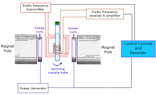
Figure1: Basic illustration of NMR
For successful use of this technique as an analyzing tool its necessary to know the physical principles of the spectrometers and methods on which the spectrometers operate. The following are the phenomenon upon which the spectrometer was developed; Charge spinning generates a magnetic field which results to spin magnetic that has a magnetic momentum that is proportional to the spin direction. Moreover, the presence of a high field of magnet originating from an external magnet creates two spins that are the +1/2 and -1/2 spins. The moment of the lower magnetic energy +1/2 gets aligned with to external magnetic field. Moment of the high spin -1/2 gets opposed to the external magnetic field. Differences in the energies between the two spin are dependent on the strength of the external field.
Strong fields of the magnet are necessary for the nuclear magnetic resonance spectroscopy to take place. NMR spectrometer is composed of three basic parts namely; computer, spectrometer, and superconducting magnet. The computer is the instrument control that runs and controls the spectrometer it also does the data processing and interpretation of the results in a language that we can understand. The spectrometer is the one that transmits the radio frequency and receives the radiofrequency. The superconducting magnet generates a strong field that is much more the earth’s magnetic field and it’s where the samples are placed and exposed to the radio waves.
From the nuclear magnetic resonance spectra, the following can be noticed, the chemical shift, relaxation time, spin-spin coupling, and the signal intensity. All these parameters are very crucial during the analysis of the sample and they give both the quantitative information, qualitative and general information about the sample analyte. With this information it’s possible to predict various reactions involving unknown substances also the purity of a certain known sample can be analyzed, the molecular dynamics and composition of the mixture can be identified.
Throughout time scientists around the globe are working on improving the spectrometer through analysis, research, and modernization of the nuclear magnetic resonance to diversify the applicability of the nuclear magnetic resonance. In general, there are two types of spectrometer, continuous- wave and the Fourier- Transform nuclear magnetic resonance. Continuous waves are one of the widely used but have been replaced with the Fourier- Transform nuclear magnetic resonance. Continuous nuclear magnetic resonance is applied mainly because its operating cost is lower compared to FT- nuclear magnetic resonance. CW- nuclear magnetic resonance is cooled with water during the low resolution while magnets in FT- nuclear magnetic resonance are cooled with liquid helium.
Discussion
Nuclear magnetic resonance technique is a technique that has been subjected to various modifications. The current and the future technologies of these techniques are key to finding solutions to most complex analysis and broadening the applicability of the technology in various industries such as food, textile, gas, and oil, medicine, etc. some of the current technologies that are being applied in nuclear magnetic resonance are;
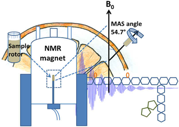
Development of Magic –Angle Spinning technology that has made the application of the solid-state chemistry in various fields. Magic Angle Spinning allows high spectra resolution in viscous samples and solids. Special situations are considered when ordinary magnetic fields at interphases are incompletely averaged out like in cases of movement of embedded molecules. In heterogeneous systems, the application of the MAS nuclear magnetic resonance has resulted in an appreciable increase in the resolution of spectra where internal motion is characterized by a relatively time correlation that is short.
Also, there has been a change in the application of the nuclear magnetic resonance spectroscopy for instance in metabolism where the collection of large amounts of spectra then gathering enough underlying data on metabolites, then it reveals the statistical analysis of the makers of various diseases or disorder. During the measurement of unknown samples, outliers from the normal data set indicate that a disease or a disorder is present and this information can be used by the clinical scientist.
Researchers in biopharmaceuticals are using nuclear magnetic resonance to characterize the structure of the monoclonal antibodies. Monoclonal antibodies are antibodies that are made when unique white blood cells are cloned. Therefore, nuclear magnetic resonance is used to characterize the structure of these antibodies. Also during the piloting of the biological drug and before full processes implementation of production scientists rely on nuclear magnetic resonance to optimize and conduct an evaluation of the nutritional growth of the media. In pharmaceutical where a small amount of fluorine is being used NMR gas also finds its way in. The use of NMR spectroscopy is used since cryoprobes can detect fluorine.
In the food industry, nuclear magnetic resonance is becoming more of an analytical tool. It is being used to screen wines to identify the country of origin of the wine and the types of grape that was used to make the wine, the consumer of the wine can be in a position to understand the type of wine he/she is consuming. This technique also has the power to determine the country of origin of the fruits that have been used to make a particular juice, and the method of the treatment the fruit underwent. For instance, it is possible to tell whether the fruit was cut before being squeezed off if it was just squeezed during the process of preparation of the juice. It is also becoming a standard tool in the diagnosis of metabolic disorders where only one spectrometer can detect more than 30 disorders and diseases. It is has found large applications in diagnostic laboratories where analytical laboratory are carried out.
In terms of the instrumentation of the NMR, several improvements have been made and are being used at the moment; multinuclear NMR during the development of NMR the studies were with highly susceptibility detection but at the moment improvements have been made and therefore studies with less susceptible nuclei are possible due to the improvements in the sensitivity of the nuclear magnetic resonance. Initially, the trend had a separate radio frequency for every nucleus, this has been changed by the improvements that have been made in that there has been the development of broadband which allows for rapid changeovers between the nucleus.
Computer developments are one of the major current changes that have occurred in relation to the technology improvements of the nuclear magnetic resonance. Modern soft wares are being used to run the spectrometer, this soft wares can handle several experiments whereby various factors that include, the time delay, widths of the pulse, decoupler levels, and the radiofrequency can be varied. The spectroscopic function is single-handedly handled by the computer. The processing capabilities that are possessed by these modern computers have expanded and enabled efficient storage and manipulation of large data and arrays.
Several methods have also been developed and improved as far as the nuclear magnetic resonance is concerned. These methods are;
Multiple Quantum nuclear magnetic resonance
Two Dimension FT nuclear magnetic resonance
High-resolution nuclear magnetic resonance
Nuclear magnetic resonance Imaging
Chemical induced dynamic polarization.
2-D FT nuclear magnetic resonance spectroscopy
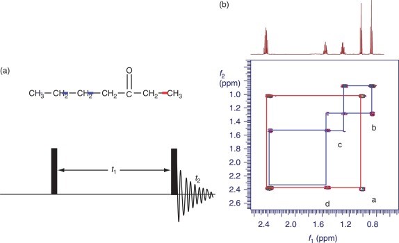
In this method, the data collection is a function with independent variables, t1, and t2 its these variable domains are followed by a 2-D Fourier Transformation.
Resulting two-dimension spectra contains two frequency axis and an intensity axis. It then becomes possible to perform several experiments.
Figure 2: Cosy pulse sequence and 2d Cosy spectrum of 3- heptanone
Multiple Quantum NMR
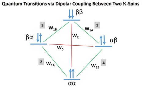
Signals that are observed from the normal nuclear magnetic resonance are from the transition that obeys the selection rules the selection rules are given by the change in m= –+1 and m represents the magnetic quantum number of the system. Using specialized techniques it’s possible to excite multiple quanta within the states of change in that m = –+2, 3, 4…. The coherences that are experienced may be converted by using the pulse techniques to signals that are detectable.
This method is widely used to provide crucial information about the motion of the compound, conformations, and molecular structure.
Figure 3: Showing multiple quantum transitions
Nuclear magnetic imaging
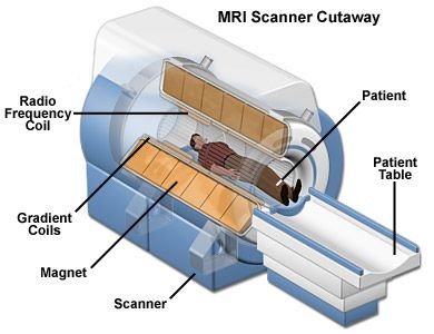
This is a non-invasive technique that enables the observation of atomic structures, physiological functions, and composition of molecular tissues. The technique is mainly applied in magnetic resonance which is based on NMR spectroscopy. Nuclear magnetic resonance imaging has been developed in a special way such that it resolves the signals developed in a different direction. Nuclear magnetic resonance encoding is accomplished by the use of magnetic fragments. The fragments can introduce spatial variation around the main magnetic field. Below is an illustration of nuclear magnetic imaging.
Figure 4: Illustration of magnetic resonance imaging
Chemical induced magnetic polarization
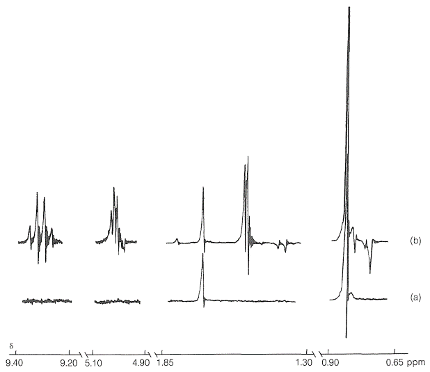
Magnetic polarization occurs by mixing the singlet and the triplets of a diffusing radical pair in a strong field of a magnet. This technique can be demonstrated by the irradiation of chemicals then observing the peaks and the intensity before and after irradiation. For instance, when 3,3- dimethyl butanone has irradiated the peaks that were shown before the irradiation according to Figure(a) the peaks were at a ratio of 3:9 but after the irradiation, the ratio changed to almost 3:18 both the intensity and the peaks.
Figure 5: below shows the peaks before and after irradiation.
Future on nuclear magnetic resonance spectroscopy
Nuclear magnetic resonance technique is continuously growing and it is expected that soon there will be many improvements would have been made for instance;
Incorporation of Artificial Intelligence analysis
Future based on metabolomics
Advancement in nuclear magnetic resonance for drug discovery
Automating nuclear magnetic resonance
The future based on metabolomics
In the field of metabolomics, nuclear magnetic resonance is still overshadowed by the mass spectroscopy MS. This is because of the number of a compound that resolved in a particular time. Nuclear magnetic resonance is highly effective at attracting pathways and fluxes. Therefore nuclear magnetic resonance has to increase the number of compounds it can resolve to match MS. With this nuclear magnetic resonance is expected to expand its amenable target.
Development of Artificial Intelligence technology in nuclear magnetic resonance
Incorporating artificial intelligence in nuclear magnetic resonance is one of the things that are most anticipated in the future. By introducing components like the descriptor of 2-D co-relation data that is associated with chemical structure will make it possible for the transfer of information to the databases.
Automating the nuclear magnetic resonance
The development of an intelligent workflow system will be an essential requirement for the efficient operation of the nuclear magnetic resonance. The new paradigms will make it possible for the aggregation of data and interactive data storage.
Advancement through drug discovery
The Discovery of drugs is complex and very unpredictable and it is associated with a high level of failure. The major contributing factors to these failures are the current trends in the fields of production that have not made it possible for ease identification and analysis of the drug. Nuclear magnetic resonance has become one of the critical components in drug production, discovery such as versatility of the nuclear magnetic resonance to ensure it can handle many targets is critical for improvement and increase in the production of drugs. Nuclear magnetic resonance technique is expected to grow in this industry with the advancement in automation, cell imaging, speed of calculation of the structure of a component, and increased sensitivity.
These are just examples of future trends that are expected in the nuclear magnetic resonance technique.
Advantages of NMR
Software advancements in nuclear magnetic resonance have made it to become widely used in various laboratories. Nuclear magnetic resonance can be used both in the field and large magnetic spectrometer laboratories.
Non-invasive
The greatest advantage that the nuclear magnetic resonance has is that it is noninvasive. During the imaging, both the biochemical and spatial information can be obtained without damaging the tissue sample. Also, there is no ionization radiation, other techniques used in vivo involves ionization of metabolism to get the signals required.
Flexibility
In Nuclear magnetic resonance there is a wide range of processes that can be investigated using the same technique for instance during the studying of metabolism in the isolated neurons cells, the technique used in the culture media, human brain, and the animal brain is the same.
Easy sample preparation
Nuclear magnetic resonance technique does not require a tedious procedure to prepare samples. Samples to be analyzed do not undergo various treatment as they do when other spectroscopic techniques are used. Also, nuclear magnetic resonance provides crucial information about the vibration of the molecular structure without interfering with them. It’s a useful technique since it has a low maintenance cost, has high throughput compared to the other techniques, and has high reproducibility.
Current and Future uses of NMR
Nuclear magnetic resonance is used to study Tautomerism. Tautomerism is a condition of chemical equilibria that involves the transfer of hydrogen atoms between the systems at the equilibria. It is an important component in biochemistry, organic synthesis, medicines, and pharmacology. When using nuclear magnetic resonance it’s possible to observe the equilibria without shifting it. Gas-phase nuclear magnetic resonance studies have been used for the study of this phenomenon, it also provides information about the chemical exchange and conformational equilibria that other experiments cannot. The spectra observed depends on the activation energy that is separating the tautomer.
Figure 6: Tautomers observed by NMR
Used in the petroleum industry
Normally in low field nuclear magnetic resonance is used in the exploration of oil to determine the T2 which is the relaxation time of distribution of the fluids in plugs. The distributors can then be examined to provide information on; permeability, size pores, and fluid index bound. It is also used in the characterization of the refined products of gasoline.
Used in medicine
Nuclear magnetic resonance is used in medicine for diagnostic purposes. In dentistry, these techniques are being used to explore several thongs like the structure of amorphous glasses, bioactive glasses, oral tissues, saliva metabolites, dental cement, and understanding of periodontal diseases.
Multiple sclerosis disease is not easily identified, nuclear magnetic resonance has since grown to use as the primary diagnostic tool.
Used in catalysis
Nuclear magnetic resonance has proven to be an important tool in accessing and understanding the catalyst surfaces of the heterogeneous catalyst. Among the technique used are the Cross polarization or Magic angle spinning. Studying the catalyst surfaces is very crucial since these surfaces are where the reactions occur. Better understanding leads to a high conversion of the reactants to products. It is therefore possible to say that nuclear magnetic resonance has made it easier for the characterization of the catalyst and understanding of the suitable catalyst and on what reactions.
Figure 7: showing magic angle spinning
Used in organic synthesis
Nuclear magnetic resonance techniques are a very reliable method of analysis and are more useful since they can be used to identify the presence of intermediate products that are formed in the course of the reactions. During the manufacture of various compounds in the industries, nuclear magnetic resonance is used to identify the structures and the arrangement of the atoms in a field of reaction. It has therefore become more useful as even the unknown samples can be detected and reaction easily predicted.
Importance of NMR in future science
Nuclear magnetic resonance is a technique that will be of most use in terms of the analysis of compounds. Nuclear magnetic resonance is a tool for authentication of food. Approaches allow specific identification by specific markers to given foodstuff, a targeted approach is used where the chemical profile of the foodstuff and beverage drinks create a unique fingerprint that is used as a reference for suspected samples of food fraud.
In cancer research, advances that are currently being made prove that the new approaches allow for early detection of tumorous cells and malignant tissues where target chemotherapy can be done.
In meat science, this spectroscopy has been used to investigate the effects of slaughter factors on meat quality through its action on phosphorous metabolism. It is also used for authentication of fresh meat by using the nuclear magnetic resonance imaging technique which is applied in the brine dynamics when meat is being processed.
Environmental science is a discipline which connects both Physico-chemical and other sciences to study problems of the environment and the impact human possesses on the environment. Solid-state nuclear magnetic resonance nuclear magnetic resonance NMR is used in the study of processes in the environment and their possible remediation, with examples from the samples collected in the field and laboratory. Other topics that are present in environmental science are phosphorus recovery, soil chemistry, and environmental remediation. Solid-state nuclear magnetic resonance technique is a strong technique used to provide atomic-level information and the interaction that occurs between pollutants and minerals on various environmental interphases. Nuclear Magnetic Resonance a tool in metal oxide cluster chemistry.
Efforts are underway for the expansion of pressure ranges for nuclear magnetic resonance in solution. As it is known, the pressure of the earth is felt greatly deep below the earth’s crust which is over forty kilometres deep. Scientists and geochemists are evaluating models of a solution of equilibrium which can be used to calculate solute activities at these very high pressures. One of the models ta has been developed is the HKF Model which employs the principle of the variable static dielectric constant of water.
Considerations of interpretation of the nuclear magnetic resonance spectrum at these elevated pressures must be taken into account. For instance, when the dielectric constant of water is over a hundred GPa, changes are non-linear.
Used in the characterization of polymers
Nuclear magnetic resonance technology is instrumental in the characterization of polymers. This is because relaxation time and chemical shifting are sensitive to the microstructure of polymer. During early stages of incorporation of this technique for characterization of polymers, high resolution of spectra was never expected because it is known that the width line depends on the third power of the rigid diameter of the sphere. This simply implied that sharp lines are associated with small molecules but not for molecules with higher molecular weight.
Spectra with high resolutions are observed in polymers because the differences between the line widths are less and hence chemical shifts and variation are due to chain chemical structure. Variation in nuclear shielding that arises from the high electron density is the main reason chemical shift resolution occurs.
The main effect of chemical shift is observed in chemical type methyl, aromatic groups, methylene, and resonance arising bring a well-separated nuclear magnetic resonance spectrum. In polymers, the spectrum is also affected by the spin of the nearby atoms. For instance, if carbon is bonded to a proton, the field that it will experience depends on the nature of alignment of the proton to the magnetic field. The carbon will split into two signal, a phenomenon that is often referred to as scalar coupling. The coupling provides information on the number of protons and other groups that are bonded to the carbon. In the process, scalar coupling makes the spectrum more complex and thus the need to suppress the coupling.
Weaknesses of Nuclear Magnetic Resonance
Nuclear magnetic resonance is less sensitive. This is because the signal that is generated by the nuclear magnetic resonance is very small for experimental purposes and depends on the concentration of the nuclei. If for instance a sample like the human body, which contains 70% water, it is hard to generate a signal since there is a low concentration of the nuclei.
It is risky working in a high magnetic field environment. Working with nuclear magnetic resonance with a high magnetic field poses a lot of risk to the personnel involved. The magnetic field can affect other equipment that is around, functioning capabilities.
Sensitivity to motion. Nuclear magnetic resonance machines are very sensitive to motions, especially during spatial encoding. This sensitivity to motion leads to distortion of the signal and malfunction of other electronic gadgets that are found.
How to overcome the nuclear magnetic resonance limitations
The versatility of nuclear magnetic resonance is one of the reasons the technique is admired but it fails mostly due to low sensitivity, this low sensitivity is due to the magnetic resonance of the nuclear magnetic resonance. The magnetic resonance has weak energy that is involved in an interaction. There are several ways in which this problem can be overcome. By optimizing the hardware used in nuclear magnetic resonance for example, the ratio between the signal and noise can be improved by increasing the concentration of analyst of interest and quicker recovery of magnet spins.
Boltzmann distribution may also be applied to enhance the signal to noise ratio through enhancing population difference. An experiment may also be carried out in static magnet fields, the static magnet field limits the usefulness of the nuclear magnetic resonance spectrum
Magnetic field drift. The drift of the field of magnet affects the resonance imaging in spectroscopy. It leads to broadening of the spectral peaks and distortion of the lineshapes. This makes interpretation of nuclear magnetic resonance very difficult. During experiments with solutions in nuclear magnetic resonance spectroscopy, it is easier to solve this problem. It is solved through correction of the field of the magnet during the recording of the measurements. Deuterium present in nuclear magnetic resonance enables tracking of the field drift when it is adjusted subsequently using room temperature electromagnet.
Magic angle spinning cannot be used to correct the magnetic drift because the samples used are only in small volumes; thus, Deuterium presence is usually very low. Currently, methods have been developed to account for the drift in the magnetic spectrum. These methods include the use of shorter blocks data acquisition that is paired with a linear calculating phase ramp and applying it in all dimensions of the spectrum. Certain algorithms have proven very useful because they are capable of correcting the drift in multiple dimensions of the spectra. Also, the algorithm may be applied in data recording as plane points or just by sampling non-uniformly Sensitivity enhancement using probe heads. The probe head is a very useful key component in nuclear magnetic resonance spectrometer. It contains coils (radiofrequency coils) tuned to a certain base frequency that detects specific nuclei of a certain magnetic field. The probeheads also have the required hardware to control temperatures. When connected to an external source of temperature, they optimize the signal-noise ratio. The geometry of probe heads is central to the performance of nuclear magnetic resonance. They are made using two coils, of which one is closest to the sample, and the other one is far away from the sample. This means that there is an outer and inner coil. These coils allow the probehead to respond to many frequencies and also allow irradiation of several nuclei. The nucleus that is in the inner coil is detected with very high sensitivity. For instance, when performing a test with a compound with carbon -13 and hydrogen-1, the inner coil would be tuned to observe the carbon-13 and the outer core will observe the hydrogen-1. Such probeheads operate in principle of polarization transfer from the highly sensitive hydrogen nucleus.
In nuclear magnetic resonance in solution, there are several specific probeheads that are of high resolution. Each of the probeheads is made in such a way that each coil is specially tuned for direct detection of the target nuclei. The probes that are used commonly are;
1H selective normal. This probehead is highly selective to the detection of hydrogen-1.
19F selective normal. The inner coil in this probehead is tuned to observe 19F.
Selective probeheads for specific nuclei. The inner coil is tuned to observe the specific nucleus.
Selective dual probe. This probe contains a decoupling channel that is designed in such a way that can be used to decouple. The probeheads are tuned to observe hydrogen- and carbon-13 nuclei
Dual probe head. They observe a certain nucleus in addition to the nucleus of hydrogen-1. It is ideal in the analysis of 31P in biofluids.
Selective indirect probes. This probeheads are made in such a way that the inner coil observes hydrogen-1 and the outer coil is tuned to observe another nucleus that is decoupled.
Broadband probes. In broadband, the inner coil follows a range of nucleus basically called the broadband channel. The channel allows coverage of large frequency. It is designed to analyze all nuclei ranging from 31P to 109Ag. However, this broadband has low sensitivity because of its non-specificity.
Conclusion
GE should re-enter the market and start manufacturing the nuclear magnetic resonance since the market is still much unexploited and has great potential in the future. Because they possess experience with nuclear magnetic resonance they have a repeatable background and should use that to their advantage.


